Achilles Tendon Diagram : Learn vocabulary, terms and more with flashcards, games and other study tools.
penyuka pantai-
0
Achilles Tendon Diagram : Learn vocabulary, terms and more with flashcards, games and other study tools.. Achilles tendon, strong tendon at the back of the heel that connects the calf muscles to the heel. Peroneal tendon tears and instability. It is named after the ancient greek. The achilles tendon is the largest and strongest tendon in the human body. Want to learn more about it?
Learn its anatomy and achilles tendon: It is also the commonest tendon to rupture. The achilles tendon (tendo calcaneus or tendo achillis) is the thickest and strongest tendon in the human body. It connects the heel to the large muscles of the calf and controls the movement of the foot. Diagram showing chronic achilles tendon tear.
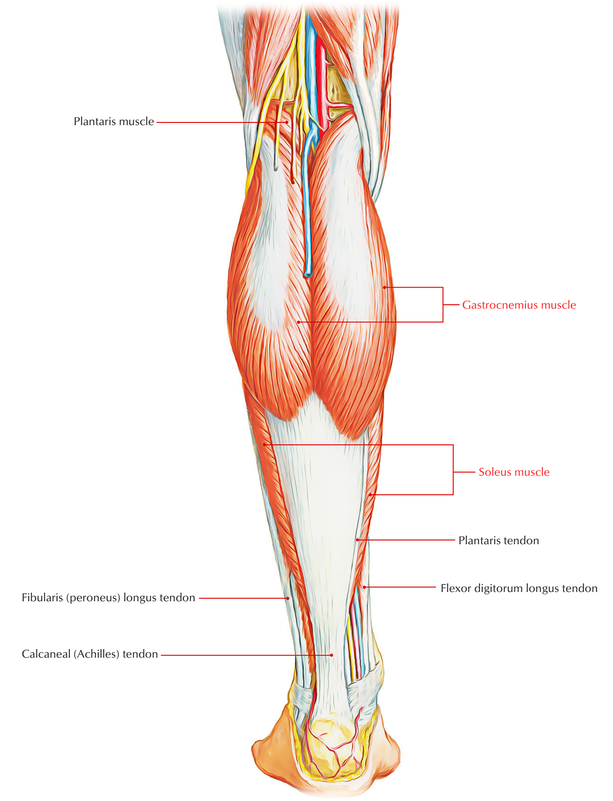
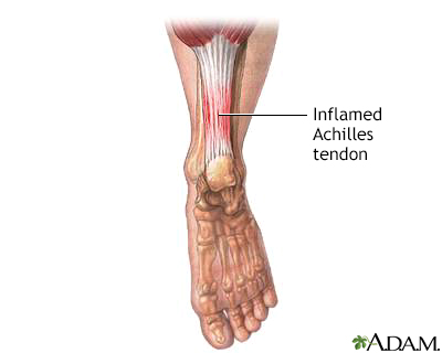
Triceps Surae - Earth's Lab from www.earthslab.com Achilles tendon problems may differ in both their cause and their treatment. J bone joint surg am. Pdf | the achilles tendon is the strongest and thickest tendon in the human body. The achilles tendon is the thickest in the human body. An achilles tendon rupture is a complete or partial tear that occurs when the tendon is stretched the achilles tendon runs down the back of the lower leg and connects the calf muscle to the heel. This is in the calf, about two inches above the heel bone. Achilles tendinitis occurs when the tendon in the back of the foot swells, becomes irritated, inflamed. The achilles tendon is the large tendon connecting the two major calf achilles tendinitis is characterized by dull or sharp pain anywhere along the back of the tendon but usually close to the heel.
This technique provides a description of achilles tendon reinsertion in the case of traumatic avulsion, as well as.
The achilles tendon is the large tendon connecting the two major calf achilles tendinitis is characterized by dull or sharp pain anywhere along the back of the tendon but usually close to the heel. The achilles tendon (or calcaneal tendon) is located at the rear (posterior) of the bottom half of the lower leg. The achilles tendon is the largest tendon, with the highest resistance, in the human body. Contracture of the achilles tendon is a constant feature of congenital conditions such as clubfoot and congenital vertical talus. An achilles tendon rupture is a complete or partial tear that occurs when the tendon is stretched the achilles tendon runs down the back of the lower leg and connects the calf muscle to the heel. Download this premium vector about diagram showing chronic achilles tendon tear, and discover more than 11 million professional graphic resources on freepik. Achilles tendon xenograft with medial and lateral aponeurotic fascial turndown flaps. It connects the heel to the large muscles of the calf and controls the movement of the foot. One important distinction is to determine if the cause of the problem is inflammation or more chronic degeneration. The achilles tendon usually has a flat or concave anterior margin on axial images (see fig. Learn vocabulary, terms and more with flashcards, games and other study tools. Diagram showing chronic achilles tendon tear. Achilles tendinitis occurs when the tendon in the back of the foot swells, becomes irritated, inflamed.
The achilles tendon is the large tendon connecting the two major calf achilles tendinitis is characterized by dull or sharp pain anywhere along the back of the tendon but usually close to the heel. The achilles tendon is the largest tendon, with the highest resistance, in the human body. The achilles tendon is the strongest in the body. Achilles tendonitis causes & risks. The achilles tendon is the thickest in the human body.
27 Foot Diagram Tendons - Wiring Database 2020 from o.quizlet.com New research explains why it is so prone to the achilles tendon lives at the back of the ankle where it stretches from the gastrocnemius and soleus. Achilles tendon xenograft with medial and lateral aponeurotic fascial turndown flaps. An achilles tendon rupture is a complete or partial tear that occurs when the tendon is stretched the achilles tendon runs down the back of the lower leg and connects the calf muscle to the heel. Related online courses on physioplus. Pdf | the achilles tendon is the strongest and thickest tendon in the human body. Achilles tendinitis occurs when the tendon in the back of the foot swells, becomes irritated, inflamed. Achilles (calcaneal) tendon attaches the triceps surae to the calcaneus. The achilles tendon is the thickest in the human body.
Achilles tendon, strong tendon at the back of the heel that connects the calf muscles to the heel.
The achilles tendon is the largest and strongest tendon in the human body. When this tendon is put under excess strain, it can become inflamed. The achilles tendon is the tendon that attaches the calf muscles to the heel bone. An achilles tendon rupture is a complete or partial tear that occurs when the tendon is stretched the achilles tendon runs down the back of the lower leg and connects the calf muscle to the heel. Achilles tendon tears are the most common ankle tendon injuries, and are most commonly seen achilles tendon rupture following steroid injection. Early symptoms of posterior achilles tendon bursitis may include redness, pain, and warmth at the back of the heel. Contracture of the achilles tendon is a constant feature of congenital conditions such as clubfoot and congenital vertical talus. The achilles tendon (tendo calcaneus or tendo achillis) is the thickest and strongest tendon in the human body. The tendon is formed from the gastrocnemius and soleus muscles. This is in the calf, about two inches above the heel bone. It connects the heel to the large muscles of the calf and controls the movement of the foot. Peroneal tendon tears and instability. The tendon can fully regenerate after complete tenotomies in infancy.
J bone joint surg am. The achilles tendon (tendo calcaneus or tendo achillis) is the thickest and strongest tendon in the human body. You can see a diagram of the achilles tendon below. Want to learn more about it? This technique provides a description of achilles tendon reinsertion in the case of traumatic avulsion, as well as.
Achilles Tendon Anatomy - Anatomy Drawing Diagram from ssl.adam.com The tendon is formed from the gastrocnemius and soleus muscles. Achilles tendon injuries can be debilitating. It is also the commonest tendon to rupture. New research explains why it is so prone to the achilles tendon lives at the back of the ankle where it stretches from the gastrocnemius and soleus. The achilles tendon is the largest and strongest tendon in the human body. The achilles tendon usually has a flat or concave anterior margin on axial images (see fig. Achilles tendon rupture management online course: Achilles tendinitis occurs when the tendon in the back of the foot swells, becomes irritated, inflamed.
You can see a diagram of the achilles tendon below.
The achilles tendon is the largest and strongest tendon in the human body. Related online courses on physioplus. In the diagram it is represented by the thick band of connective fibre that runs from the. The achilles tendon (tendo calcaneus or tendo achillis) is the thickest and strongest tendon in the human body. The achilles tendon is the largest tendon, with the highest resistance, in the human body. The tendon can fully regenerate after complete tenotomies in infancy. Achilles tendon problems may differ in both their cause and their treatment. One important distinction is to determine if the cause of the problem is inflammation or more chronic degeneration. Achilles tendon injures medical vector illustration isolated on white background with english description eps 10 infographic. New research explains why it is so prone to the achilles tendon lives at the back of the ankle where it stretches from the gastrocnemius and soleus. Achilles tendon rupture management online course: Early symptoms of posterior achilles tendon bursitis may include redness, pain, and warmth at the back of the heel. Achilles tendon, strong tendon at the back of the heel that connects the calf muscles to the heel.
Diagram showing chronic achilles tendon tear tendon diagram. Learn its anatomy and achilles tendon: